
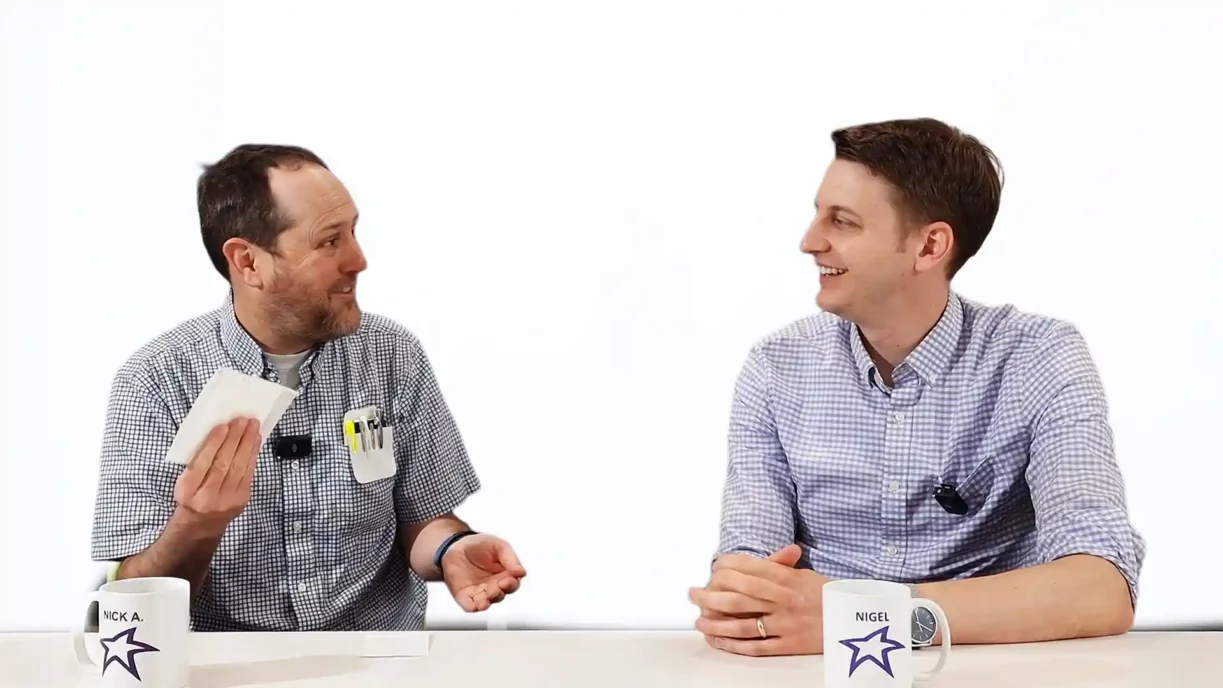
Sandwich ELISA Assay Explained

The sandwich ELISA assay is one of the most common ELISA formats used in diagnostics. In this Bio Break episode, Nick and Nigel walk through the method step by step using simple visuals and plain language. As a result, the concept becomes much easier to understand, even if immunoassays are not your day job.
At its core, a sandwich ELISA assay uses multiple antibodies to detect a single target antigen. The name comes from how the antigen is captured between two antibodies, much like the filling between slices of bread. This structure allows for strong binding, reliable washing, and clear signal generation.
Capturing the Antigen
First, an antibody is fixed to the surface of an ELISA plate. This antibody is designed to recognize a specific antigen, such as a virus or other biological marker. When a sample flows across the plate, the antigen binds to this anchored antibody. Anything that does not bind can then be washed away.
Because of this initial capture step, the target stays firmly attached to the plate. That stability is important, since ELISA relies on multiple wash steps to reduce noise and improve accuracy.
Building the Antibody Sandwich
Next, a second antibody is introduced. This antibody recognizes a different site on the same antigen. As a result, the antigen becomes bound between two antibodies. This creates the sandwich structure that gives the sandwich ELISA assay its name.
However, the detection does not stop there. A third antibody is added that recognizes the second antibody. This third antibody carries an enzyme. When a substrate is added, the enzyme reacts and produces a detectable signal. At that point, the presence of the antigen becomes visible or measurable.
Why This ELISA Method Is So Effective
Because the antigen must be recognized by multiple antibodies, the sandwich ELISA assay offers strong specificity. In addition, the washing steps remove unbound material, which helps improve signal clarity. For these reasons, this method is widely used in laboratory testing and diagnostic workflows.
Enjoying Bio Break? Sign up to get new episodes sent to your inbox.
Related Resources

Nigel Syrotuck breaks down REACH SVHC compliance for teams working with material suppliers and compliance questionnaires.

We examine when computational modelling and simulation, or CM&S, genuinely supports medical device simulation strategy and when it becomes a costly detour.
Many teams still underuse CM&S, often bringing it late in device validation, when key decisions have already been made. That approach leaves much of the value of CM&S untapped.

This article traces the Pennes bioheat equation from its 1948 origins to modern multiscale approaches, explaining how engineers select the right level of modelling complexity across device categories.